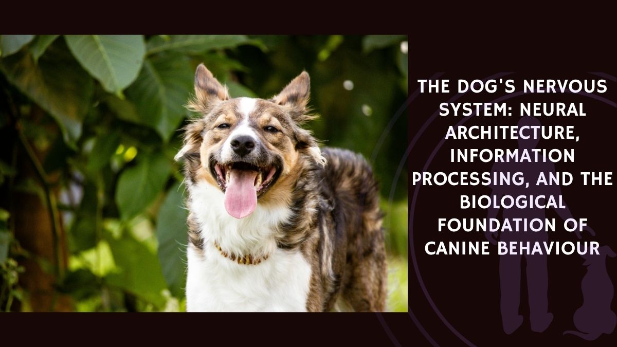
Hippocampal atrophy is a hallmark feature of human Alzheimer’s disease (AD), visible both pathologically and through magnetic resonance imaging (MRI). Until recently, this phenomenon had not been documented in dogs with canine cognitive dysfunction (CCD), a natural model of human dementia. Dewey and colleagues (2020) addressed this gap through a retrospective comparative MRI study.
The research team analyzed MRI scans from 42 aging dogs (≥9 years old), including 16 CCD-diagnosed dogs and 26 successfully aging controls. Using Mimics® software, they calculated total hippocampal and brain volumes. The study found that hippocampal volume normalized to total brain volume was significantly smaller in CCD dogs compared to controls (p = 0.04).
These findings suggest that hippocampal atrophy is a pathological feature of CCD, closely mirroring human AD. This structural change underscores the value of dogs as a model for studying dementia and suggests that hippocampal-targeted therapies—already explored in Alzheimer’s research—may also hold promise for canine patients.
By identifying hippocampal shrinkage as a shared biomarker of neurodegeneration across species, this study strengthens the translational potential of CCD research and may help guide both veterinary and human treatment strategies.
Source: Dewey, C. W., Rishniw, M., Platt, S., Robinson, K. R., Sackman, J., & O’Donnell, M. (2020). Canine cognitive dysfunction (CCD) patients have reduced total hippocampal volume compared with aging control dogs: a comparative MRI study. bioRxiv, published January 27, 2020.