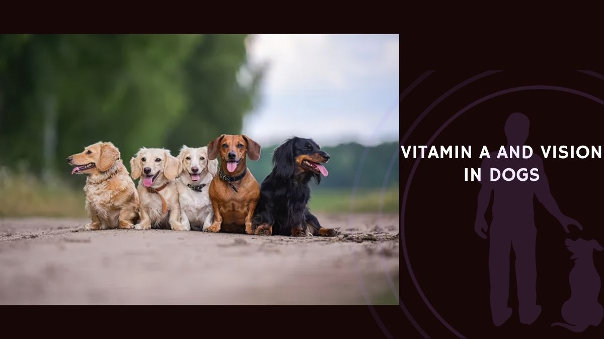
Your dog’s eyes are extraordinary. Designed for twilight hunts and wide-angle awareness, they rely on a single nutrient more than almost any other to keep the lights on — literally. That nutrient is vitamin A. Without it, the intricate machinery of your dog’s retina grinds to a halt, the protective surface of the eye dries and cracks, and the immune defenses that guard every mucous membrane begin to fail.
But here is the twist most owners never hear: too much vitamin A is just as dangerous as too little, and the line between “enough” and “too much” is narrower than you might think.
In this guide, we walk through everything the research tells us about vitamin A and canine vision — from the molecular events inside a single photoreceptor cell all the way to the bowl of food you set down tonight. Every finding, every mechanism, every clinical sign. Because understanding the full picture is the only way to make truly informed decisions for your dog’s eye health. 🐾
Vitamin A Biology: What It Is, Where It Comes From, and How Your Dog Uses It
Dietary Sources and Absorption
Vitamin A cannot be manufactured inside your dog’s body. It must come from the diet, and it arrives in two distinct forms.
Preformed vitamin A (retinol) comes from animal-based ingredients and is the most efficiently absorbed form. The richest sources include:
- Liver and organ meats (by far the highest concentration)
- Egg yolks
- Fish and fish oils (particularly cod liver oil)
- Milk and dairy products (in moderation)
- Salmon and other fatty fish
Provitamin A carotenoids, primarily β-carotene, come from plant-based ingredients. Good sources include:
- Carrots, sweet potatoes, and winter squash
- Spinach and other dark leafy greens
- Pumpkin
- Broccoli
- Red bell peppers
- Oranges, mangoes, cantaloupe, and watermelon
Here is an important distinction: dogs absorb preformed retinol far more efficiently than they convert β-carotene into active vitamin A. The conversion of carotenoids to retinol is variable and significantly less efficient than direct absorption of preformed retinoids. This means that plant-based sources alone may not reliably meet all of your dog’s vitamin A requirements — a fact that matters enormously for owners feeding homemade or vegetable-heavy diets.
Practical Portions: How Much Vitamin A Is Actually in Your Dog’s Food?
Knowing that liver is “high in vitamin A” is not enough. Owners who prepare homemade diets or add whole-food toppers need real numbers to avoid both deficiency and toxicity. The following approximate values will give you a sense of scale.
Animal-based sources (preformed retinol, approximate IU per portion):
- Beef liver, 30 g (about 1 oz): ~8,000–9,000 IU
- Chicken liver, 30 g (about 1 oz): ~4,000–4,500 IU
- Cod liver oil, 1 teaspoon (5 ml): ~4,000–5,000 IU
- Whole egg, 1 large: ~250–300 IU
- Salmon, 85 g (3 oz) cooked: ~50–60 IU
- Whole milk, 100 ml: ~40–50 IU
Plant-based sources (provitamin A carotenoids, approximate IU equivalent per portion):
- Sweet potato, 100 g baked with skin: ~19,000 IU equivalent
- Carrot, 1 medium raw (~60 g): ~5,000–6,000 IU equivalent
- Pumpkin, 100 g cooked/canned: ~6,000–7,000 IU equivalent
- Spinach, 50 g cooked: ~4,500–5,000 IU equivalent
- Red bell pepper, 1/2 medium: ~1,500–1,800 IU equivalent
- Broccoli, 80 g cooked: ~600–800 IU equivalent
- Mango, 100 g: ~1,000–1,200 IU equivalent
- Cantaloupe, 100 g: ~3,000–3,400 IU equivalent
A critical caveat with the plant-based numbers: these values represent the total carotenoid content expressed in IU equivalents, but your dog’s actual conversion of β-carotene to usable retinol is significantly lower than the raw numbers suggest. Dogs convert carotenoids less efficiently than the theoretical maximum, so a sweet potato delivering 19,000 IU equivalent of β-carotene does not provide the same usable vitamin A as 19,000 IU of preformed retinol from liver. This is why animal-sourced vitamin A remains the most reliable form for meeting canine requirements.
Look at those liver numbers closely. Just 30 grams of beef liver — barely a mouthful for a medium-sized dog — delivers roughly 8,000–9,000 IU. If your dog is already eating a complete commercial diet formulated to meet vitamin A requirements, adding liver as a daily “treat” or topper quickly pushes cumulative intake into dangerous territory over time. Occasional liver as a small treat is one thing; daily liver as a meal component on top of a complete diet is a very different calculation. 🧠
Transport: How Vitamin A Gets Where It Needs to Go
Once your dog absorbs dietary retinol, it undergoes a complex journey through the body before it can do its work.
In the intestinal lining, retinol is esterified (chemically packaged) and loaded onto transport particles called chylomicrons, which carry it through the bloodstream to the liver. The liver is the central warehouse. There, retinol is either stored for later use or re-esterified and kept in reserve.
When the body needs vitamin A, the liver releases retinol bound to a specialized carrier called retinol-binding protein 4 (RBP4). This RBP4-retinol complex circulates in the blood and delivers vitamin A to the tissues that need it.
Two key membrane receptors control how cells pick up circulating RBP4-retinol:
- STRA6 (Stimulated by Retinoic Acid 6) — operates primarily in the eye and other peripheral tissues
- RBPR2 (Retinol Binding Protein Receptor 2) — handles uptake in the liver and systemic tissues
These receptors bind the circulating RBP4 complex and pull retinol into the cell — a process that is absolutely critical for maintaining the right balance of retinoids in the eye and throughout the body.
Inside the Cell: Activation and Feedback
Once retinol enters a cell, it undergoes enzymatic conversion into its biologically active forms.
Retinal (retinaldehyde) is produced when retinol is oxidized by alcohol dehydrogenases and retinol dehydrogenases. Retinal is the molecule that directly participates in vision — it is the light-sensitive component of the visual pigments in your dog’s retina. It also serves as the precursor for the next metabolite.
Retinoic acid (RA) forms when retinal is further oxidized by aldehyde dehydrogenases. Retinoic acid is the primary signaling form of vitamin A. It enters the cell nucleus, binds to retinoic acid receptors (RARs) and retinoid X receptors (RXRs), and directly regulates gene expression. This is how vitamin A influences cellular differentiation, immune function, and tissue maintenance at the deepest molecular level.
Feedback regulation keeps all of this in check. When retinoic acid levels rise, the body suppresses the enzymes that produce more retinoids and ramps up the enzymes that break retinoic acid down. This built-in thermostat protects against both deficiency and toxic accumulation. It is elegant biology — but it has limits, which is why chronic oversupplementation can overwhelm the system.
The Liver: Your Dog’s Vitamin A Vault
The liver is the primary storage organ for vitamin A in dogs. Retinyl esters accumulate inside specialized hepatic stellate cells, creating a substantial reserve that can sustain normal retinoid function for weeks to months, even during periods when the diet falls short.
This storage capacity has two practical consequences. First, it explains why acute vitamin A deficiency is relatively uncommon in dogs eating varied diets — there is a buffer. Second, and less reassuring, it explains why chronic excessive supplementation can quietly build up to toxic levels in the liver long before any outward signs appear.
The Visual Cycle: How Vitamin A Powers Your Dog’s Sight
Rhodopsin and the Miracle of Phototransduction
The fundamental role of vitamin A in vision centers on its participation in the visual cycle — the biochemical process by which photons of light are converted into neural signals your dog’s brain can interpret.
At the heart of this process sits rhodopsin, a G-protein-coupled receptor embedded in the outer segment membranes of rod photoreceptors. Rhodopsin consists of the protein opsin covalently bound to 11-cis-retinal — the aldehyde form of vitamin A. This molecular partnership is what makes vision possible.
When light enters the eye and strikes the retina, photons are absorbed by 11-cis-retinal. The energy of the photon causes 11-cis-retinal to change shape, isomerizing to all-trans-retinal. That tiny conformational shift triggers a cascade of intracellular signaling events that ultimately hyperpolarize the photoreceptor cell and generate a neural signal transmitted to the brain. This is the moment of seeing.
But here is the challenge: once 11-cis-retinal has absorbed a photon and flipped to the all-trans form, it must be regenerated before it can detect another photon. This regeneration happens through a precisely ordered series of enzymatic steps known as the visual cycle:
- All-trans-retinal is reduced to all-trans-retinol by retinol dehydrogenase
- All-trans-retinol is transported from the photoreceptor to the retinal pigment epithelium (RPE)
- In the RPE, all-trans-retinol is esterified and isomerized to 11-cis-retinyl ester
- 11-cis-retinyl ester is hydrolyzed to 11-cis-retinol
- 11-cis-retinol is oxidized back to 11-cis-retinal
- 11-cis-retinal is transported back to the photoreceptors and recombines with opsin to regenerate rhodopsin
This cycle runs continuously during visual function and is absolutely dependent on an adequate supply of vitamin A. No vitamin A, no 11-cis-retinal. No 11-cis-retinal, no rhodopsin. No rhodopsin, no vision in low light.
Dark Adaptation and Low-Light Vision
One of the most clinically significant functions of vitamin A is its role in dark adaptation — the process by which the eye becomes increasingly sensitive to light in dim conditions.
Dark adaptation depends on the regeneration of rhodopsin in rod photoreceptors. Rods are exquisitely sensitive to light and are responsible for vision in low-light conditions. When vitamin A is adequate, the visual cycle operates efficiently, allowing rapid regeneration of rhodopsin and quick restoration of rod sensitivity after light exposure.
When vitamin A is deficient, the story changes. One of the earliest clinical signs of vitamin A deficiency is night blindness (nyctalopia) — difficulty navigating in dim light or darkness. This occurs because insufficient vitamin A limits the rate at which rhodopsin can be regenerated, leaving rods unable to achieve full sensitivity.
Behavioral signs that may suggest night blindness in dogs include:
- Reluctance to move or walk in dim environments
- Hesitation or freezing at doorways after dark
- Bumping into furniture or objects in low light
- Apparent disorientation when the lights go down
- Anxiety or whining during nighttime walks
- Reluctance to go outside at dusk or after sunset
If your dog seems fine during the day but becomes anxious or clumsy after dark, vitamin A status is one factor your veterinarian should investigate — though there are many other possible causes as well. 🧡
Cone Function and Color Vision
While rods depend on 11-cis-retinal for rhodopsin formation, cone photoreceptors also require retinal for their visual pigments (cone opsins). Cones are responsible for color vision and high-acuity vision in bright light.
Although cones are less dramatically affected than rods in early vitamin A deficiency, severe and prolonged deficiency does impair cone function as well — meaning that not only low-light vision but also daytime visual sharpness and color perception deteriorate when vitamin A is critically lacking.
The Retinal Pigment Epithelium: Unsung Hero of Eye Health
The retinal pigment epithelium (RPE) is a specialized layer of cells that lies between the photoreceptors and the choroid (the blood-rich layer behind the retina). The RPE performs multiple vitamin A-dependent functions that are essential for maintaining retinal health:
- Regeneration of 11-cis-retinal through the visual cycle
- Phagocytosis (ingesting and recycling) of shed photoreceptor outer segment membranes
- Transport of nutrients from the blood to the photoreceptors
- Maintenance of the blood-retinal barrier that protects the retina from harmful substances
- Antioxidant defense against oxidative stress
An interesting side note from emerging research: while not directly a vitamin A function, vitamin D deficiency has been shown to exacerbate oxidative damage and inflammation in RPE cells. Studies have demonstrated that vitamin D exposure can counteract hydrogen peroxide-induced oxidative damage and reduce pro-inflammatory cytokine production in RPE cells. This suggests that eye health depends on a network of micronutrients working together, not just vitamin A in isolation.
Ocular Surface Health: How Vitamin A Protects the Front of the Eye
Epithelial Differentiation and Maintenance
Vitamin A, particularly in its retinoic acid form, plays a crucial role in the differentiation and maintenance of epithelial tissues throughout the body — and the eye’s surface is no exception.
Retinoic acid binds to nuclear receptors (RARs and RXRs) and regulates the expression of genes involved in epithelial cell differentiation, mucin production, and barrier function. When vitamin A is adequate, epithelial cells maintain their specialized functions and structural integrity.
Ocular surface tissues that depend on vitamin A include:
- Conjunctival epithelium — the membrane lining the eyelids and covering the white of the eye
- Corneal epithelium — the transparent front surface of the eye
- Lacrimal gland epithelium — responsible for tear production
- Meibomian gland epithelium — produces the lipid layer of the tear film that prevents tears from evaporating too quickly
Mucus Production and the Tear Film
One of the most important vitamin A-dependent functions at the ocular surface is the production of protective mucus and tears.
Vitamin A deficiency impairs the differentiation of goblet cells and other mucus-secreting cells. The consequences cascade through the entire tear system:
- Reduced tear production
- Abnormal tear composition
- Loss of the protective mucus layer on the cornea and conjunctiva
- Increased susceptibility to corneal drying (xerosis)
In clinical terms, dogs with vitamin A deficiency may develop:
- Dry eyes (xerophthalmia)
- Corneal cloudiness or opacity
- Conjunctival inflammation
- Increased susceptibility to corneal ulceration
- Secondary bacterial or fungal infections of the ocular surface
These signs can appear gradually and may initially be mistaken for allergies or minor irritation before the underlying nutritional cause is identified.
Nutritional vs. Non-Nutritional Eye Disease: Getting the Diagnosis Right
This is a critical point that every dog owner should understand: many ocular conditions in dogs have nothing to do with vitamin A and should not be attributed to deficiency or excess.
Inherited retinal diseases (genetic in origin):
- Progressive retinal atrophy (PRA)
- Collie eye anomaly
- Retinal dysplasia
- Various breed-specific retinal conditions
No amount of vitamin A supplementation can prevent or treat these conditions.
Acquired non-nutritional disorders (the majority of eye problems in veterinary practice):
- Cataracts (age-related, diabetic, traumatic, or hereditary)
- Glaucoma (primary or secondary)
- Retinal detachment
- Optic neuritis
- Uveitis (infectious or immune-mediated)
- Corneal ulceration (traumatic or infectious)
- Immune-mediated keratoconjunctivitis sicca (dry eye from immune attack on tear glands)
Neurological causes of visual impairment:
- Optic nerve hypoplasia or atrophy
- Brain tumors or lesions affecting the visual cortex
- Seizure disorders that interfere with visual processing
A thorough ophthalmological examination — including fundoscopy, tonometry, and potentially imaging — is necessary to differentiate nutritional from non-nutritional causes of visual dysfunction. Reaching for a vitamin A supplement without a proper diagnosis is not just unhelpful; it can delay identification of the real problem. 🧠
Vitamin A Deficiency in Dogs: Causes, Signs, and Diagnosis
How Common Is It, and Who Is at Risk?
True vitamin A deficiency is relatively uncommon in dogs consuming commercial complete and balanced diets, because these products are specifically formulated to meet established nutritional requirements. However, deficiency can and does occur in specific circumstances.
High-risk scenarios include:
- Dogs consuming exclusively homemade diets without veterinary nutritional guidance
- Dogs with malabsorption disorders (pancreatic insufficiency, inflammatory bowel disease, bacterial overgrowth)
- Dogs with genetic mutations affecting retinoid metabolism or transport
- Dogs receiving medications that interfere with fat absorption
- Puppies born to vitamin A-deficient mothers
- Dogs with severe liver disease (impaired storage and metabolism)
- Dogs consuming diets very low in fat (vitamin A requires dietary fat for absorption)
If any of these situations applies to your dog, a conversation with your veterinarian about vitamin A status is warranted.
Clinical Signs: What to Watch For
The clinical manifestations of vitamin A deficiency in dogs reflect the vitamin’s many physiological roles. They tend to appear gradually and across multiple body systems.
Ocular signs (often among the first to appear):
- Night blindness — difficulty navigating in dim light, reluctance to move at night
- Dry eyes (xerophthalmia)
- Corneal cloudiness or opacity
- Conjunctival inflammation
- Increased susceptibility to corneal ulceration
- Abnormal or sluggish pupillary light reflex
Dermatological signs:
- Sparse fur or patchy hair loss (alopecia)
- Dry, scaly skin with bumps or lesions
- Rough or dull hair coat
- Increased susceptibility to secondary bacterial or yeast skin infections
- Unpleasant odor from infected skin lesions
Systemic signs:
- Weakness and lethargy
- Poor appetite or refusal to eat
- Weight loss
- Stunted growth (particularly in puppies)
- Increased susceptibility to respiratory infections (impaired mucus production in lungs)
- Pneumonia or recurrent respiratory infections
Developmental and reproductive signs:
- Fetal deformities in pregnant dogs (cleft palate, skeletal abnormalities)
- Impaired bone growth and development in puppies
- Abnormalities of inner ear bone structure, potentially leading to deafness or hearing loss
Immune dysfunction:
- Increased susceptibility to infections
- Impaired wound healing
- Reduced immune response to vaccination
From Subtle to Severe: How Deficiency Progresses
The severity and progression depend on how much vitamin A is missing and for how long.
Acute severe deficiency is rare in dogs but can occur if vitamin A sources are suddenly and completely eliminated. Signs may progress rapidly.
Chronic mild-to-moderate deficiency is more commonly seen in dogs eating unbalanced homemade diets. Clinical signs develop gradually over weeks to months, often beginning with subtle changes in coat quality or night vision before progressing to more obvious systemic problems.
Subclinical deficiency is the trickiest category. Vitamin A levels may be below optimal but not yet producing obvious clinical signs. This state may be detectable through blood testing but may not yet warrant intervention in an otherwise healthy, asymptomatic dog.
How Veterinarians Diagnose Vitamin A Deficiency
Clinical history and examination are the starting point. A thorough dietary history is essential. Dogs consuming exclusively homemade diets — particularly those lacking liver or other vitamin A-rich ingredients — are at higher risk.
Blood testing can measure serum retinol levels. Normal serum retinol in dogs typically falls between 20 and 100 μg/dL, though reference ranges vary by laboratory. However, serum retinol reflects recent dietary intake and hepatic stores and may not accurately capture tissue-level vitamin A status in every case.
Ophthalmological examination through fundoscopy may reveal retinal changes, though these are often subtle in early deficiency. Electroretinography (ERG) offers a more objective assessment — it directly measures photoreceptor function and may show reduced responses when vitamin A is lacking.
Imaging with X-rays may reveal skeletal abnormalities in severe deficiency, particularly in growing puppies whose bone development has been compromised.
Vision. Needs. Balance.
Vitamin A keeps your dog’s visual system working. It supports retinal function, protects the eye’s surface, and helps maintain the immune defenses surrounding delicate ocular tissues.
More is never automatically better. Dogs rely on dietary vitamin A, but excessive intake—particularly from concentrated sources such as liver—can become harmful, making nutritional balance essential.



Smart nutrition protects what matters. Understanding the difference between preformed retinol and plant-derived carotenoids helps owners make informed choices that support healthy vision without creating unnecessary risk.🐾
Vitamin A Toxicity: When More Becomes Dangerous
How Toxicity Develops
Unlike water-soluble vitamins that the body can readily excrete, vitamin A is fat-soluble and accumulates in the body — especially in the liver. When dietary intake chronically exceeds requirements, retinyl ester accumulation occurs, pushing serum retinol levels and tissue retinoid concentrations beyond what the body can handle.
At the cellular level, excessive retinoic acid dysregulates gene expression, leading to abnormal cellular differentiation, uncontrolled proliferation, and programmed cell death (apoptosis). This is particularly problematic in tissues that are actively remodeling, such as bone — which explains why skeletal abnormalities are among the hallmark signs of chronic vitamin A toxicity.
Clinical Signs of Too Much Vitamin A
Acute toxicity signs (single large dose ingestion):
- Vomiting
- Drowsiness or lethargy
- Irritability
- Peeling or desquamation of the skin
- Abdominal pain
Chronic toxicity signs (prolonged excessive intake over weeks to months):
- Poor hair coat
- Rough or dry skin
- Weakness and lethargy
- Weight loss
- Constipation
- Excessive bone development (hyperostosis), particularly in the neck and chest
- Painful or limited joint movement and stiffness
- Lameness or reluctance to move
- Abnormal bone growth and remodeling
These skeletal changes are often the finding that finally leads to a diagnosis.
Reproductive and developmental toxicity:
- Cleft palate formation
- Other fetal abnormalities
- Skeletal deformities in offspring
Both deficiency and excess during pregnancy cause birth defects — the window of safe intake is real and matters.
Severe toxicity (life-threatening):
- Seizures
- Paralysis
- Cardiac arrhythmias
- Cardiac arrest and death
Where Does the Excess Come From?
The most common sources of vitamin A toxicity in dogs include:
- Raw liver fed in large quantities or as a primary protein source
- Cod liver oil supplements given in excessive amounts
- Multivitamin supplements containing high-dose vitamin A
- Retinol-containing human skincare products (serums, creams, and retinoids a dog might lick or ingest)
- Excessive supplementation of vitamin A-rich foods (liver, sweet potatoes, carrots) added on top of an already nutritionally complete commercial diet
An important number to keep in mind: dogs can safely ingest up to 100 times the recommended daily intake of vitamin A without immediate acute toxicity. That sounds like a huge safety margin, and for a single exposure it is. But chronic intake at even moderately elevated levels — 10 to 20 times the daily requirement — can lead to cumulative toxicity over weeks to months. The margin for chronic safety is much narrower than for acute safety.
👁️🐾 Vitamin A & Vision in Dogs
From retinal biochemistry to the food bowl — everything science tells us about protecting your dog’s eyesight through smarter vitamin A nutrition
Phase 1: Vitamin A Biology
Sources, Absorption & Cellular MetabolismVitamin A arrives in two dietary forms: preformed retinol from animal sources (liver, egg yolks, fish oils) and provitamin A carotenoids (β-carotene) from plants (carrots, sweet potatoes, pumpkin). Dogs absorb preformed retinol far more efficiently — plant sources alone may not reliably meet all requirements.
• Retinol travels via chylomicrons to the liver for storage in hepatic stellate cells
• Released bound to RBP4 protein when tissues need it
• Receptors STRA6 (eyes) and RBPR2 (liver) control cellular uptake
• Converted intracellularly to retinal (vision) and retinoic acid (gene regulation)
• Feedback regulation prevents both deficiency and toxic accumulation
Hepatic reserves can sustain function for weeks to months during dietary gaps — but this same storage capacity means chronic oversupplementation builds up silently to toxic levels long before outward signs appear.
Phase 2: The Visual Cycle
Rhodopsin, Phototransduction & Dark AdaptationRhodopsin = opsin protein + 11-cis-retinal (vitamin A). When a photon hits 11-cis-retinal, it flips to all-trans-retinal, triggering a neural signal to the brain. This is the molecular moment of seeing. The spent retinal must be regenerated through a continuous 6-step enzymatic cycle — entirely dependent on adequate vitamin A supply.
• Rod photoreceptors (low-light vision) depend on rapid rhodopsin regeneration
• Deficiency slows regeneration → rods cannot achieve full sensitivity
• Dogs may hesitate at doorways after dark, bump into objects, or seem disoriented
• Cone function (color/daylight vision) also deteriorates in severe deficiency
This specialized cell layer regenerates 11-cis-retinal, recycles photoreceptor membranes, transports nutrients, maintains the blood-retinal barrier, and provides antioxidant defense. Emerging research shows vitamin D deficiency also damages RPE cells — eye health requires multiple micronutrients working together.
Phase 3: Ocular Surface Protection
Tear Film, Epithelial Integrity & Corneal Health• Conjunctival epithelium — lines eyelids and covers the white of the eye
• Corneal epithelium — the transparent front surface
• Lacrimal gland epithelium — produces tears
• Meibomian gland epithelium — produces the lipid layer preventing tear evaporation
Deficiency impairs goblet cell differentiation → reduced tears, abnormal composition, lost mucus layer. Clinical signs include dry eyes (xerophthalmia), corneal cloudiness, conjunctival inflammation, corneal ulceration, and secondary infections. Often mistaken for allergies before the nutritional cause is identified.
Phase 4: Deficiency
Risk Factors, Clinical Signs & Diagnosis• Homemade diets without veterinary nutritional guidance
• Malabsorption disorders (pancreatic insufficiency, IBD, bacterial overgrowth)
• Genetic mutations affecting retinoid metabolism
• Medications interfering with fat absorption
• Puppies born to deficient mothers
• Severe liver disease or very low-fat diets
• Eyes: Night blindness, dry eyes, corneal cloudiness, sluggish pupil response
• Skin: Patchy hair loss, scaly lesions, dull coat, secondary infections
• Systemic: Lethargy, weight loss, stunted growth, respiratory infections
• Reproductive: Fetal deformities, impaired bone development, potential hearing loss
• Immune: Increased infections, impaired wound healing, reduced vaccine response
• Blood testing: Normal serum retinol = 20–100 μg/dL
• Electroretinography (ERG): Objectively measures photoreceptor function
• Fundoscopy: May reveal subtle retinal changes
• X-rays: Skeletal abnormalities in severe cases (especially puppies)
Phase 5: Toxicity
Mechanisms, Signs & Irreversible Consequences• Raw liver fed daily or as primary protein source
• Cod liver oil given in excessive amounts
• High-dose multivitamins or human supplements
• Retinol-containing skincare products ingested by the dog
• Stacking vitamin A foods on top of a complete commercial diet
Acute (single large dose): Vomiting, drowsiness, skin peeling, abdominal pain
Chronic (weeks to months): Hyperostosis (excess bone growth) in neck and chest, joint stiffness and pain, lameness, weight loss, constipation, rough coat. Bone changes are permanent and irreversible.
Severe: Seizures, paralysis, cardiac arrhythmias, cardiac arrest
Primary treatment is dietary correction: stop the source, switch to balanced commercial food. Serum levels normalize within weeks — but skeletal changes are permanent. Elevated serum retinol can persist for years as the liver mobilizes stored reserves. Long-term pain management may be necessary.
Phase 6: The Fat-Soluble Vitamin Network
How Vitamins A, D, E & K InteractAll four fat-soluble vitamins (A, D, E, K) share absorption pathways and compete for micellar transport in the gut. When one is present in excess, it can reduce absorption of the others. They are a network — overloading one node disrupts the entire balance.
• A ↔ D: Both share the RXR nuclear receptor — excess vitamin A outcompetes vitamin D signaling, even if D intake is adequate
• A ↔ E: High-dose vitamin A can deplete vitamin E stores, reducing antioxidant protection
• A ↔ K: Excessive vitamin A interferes with vitamin K-dependent bone metabolism and clotting — compounding the skeletal damage from A toxicity
Cod liver oil delivers very high concentrations of both vitamin A and vitamin D simultaneously — making it particularly easy to overdose both nutrients at once. Never supplement multiple fat-soluble vitamins independently without veterinary guidance.
Phase 7: Breed Sensitivities & Senior Dogs
Vitamin A-Responsive Dermatosis & Aging EyesSome breeds develop follicular hyperkeratosis, scaly plaques, and waxy ear buildup that responds specifically to therapeutic vitamin A — even with normal serum levels. Most commonly affected: Cocker Spaniels (classic breed), Miniature Schnauzers, Labrador Retrievers, Shar-Peis, West Highland White Terriers, and Cairn Terriers. Requires dermatologist-supervised supplementation.
• Reduced intestinal absorption efficiency
• Altered liver function and enzyme activity
• Decreased bile production (less micellar formation)
• RPE accumulates lipofuscin waste, impairing the visual cycle
• Tear film becomes less stable → increased dry eye vulnerability
• Choroidal blood flow decreases → reduced nutrient delivery to retina
Switch to age-appropriate senior formulas. Schedule regular wellness checks and ophthalmological exams. Don’t assume vision changes are “just aging” — some have a nutritional component that is identifiable and addressable. Monitor, don’t over-supplement: seniors face elevated risk of both deficiency (reduced absorption) and toxicity (reduced hepatic clearance) simultaneously.
Phase 8: Smart Supplementation
When It Helps, When It Hurts & The 6-Step Protocol• Documented clinical or biochemical deficiency
• Malabsorption disorders (pancreatic insufficiency, IBD)
• Specific ocular conditions with confirmed or suspected deficiency
• Veterinary prescription based on individual assessment
• Dogs eating complete commercial diets (already formulated for requirements)
• Speculative “vision enhancement” — no evidence supports improved eyesight in adequately nourished dogs
• Unproven cognitive or behavioral claims
• Adding supplements or liver on top of a complete diet = unnecessary toxicity risk
1. Baseline blood testing before starting
2. Dog-specific formulations only (never human supplements)
3. Follow veterinary dosing precisely
4. Periodic monitoring to prevent toxic accumulation
5. Coordinate with overall diet composition
6. Never stack sources (supplements + liver + fortified treats = danger)
🔍 Vitamin A Needs by Life Stage & Situation
Requirements: Higher relative to body weight due to rapid growth. Risks: Most vulnerable to deficiency → stunted growth, skeletal abnormalities, impaired immune development. Deficiency effects may be irreversible.
Requirements: 3,333–5,000 IU/kg dry matter diet. Key fact: Complete commercial diets meet this. Supplementation on top creates cumulative toxicity risk. Focus on balanced diet, not extra vitamin A.
Requirements: Increased for fetal development. Critical: Both deficiency AND excess cause birth defects (cleft palate, skeletal deformities). The safe window is narrow — veterinary guidance essential.
Challenge: Reduced absorption AND reduced hepatic clearance — elevated risk on both sides. Strategy: Age-appropriate diet formulas, regular wellness checks, ophthalmological screening. Monitor, don’t blindly supplement.
Conditions: Pancreatic insufficiency, IBD, bacterial overgrowth. Impact: Reduced fat absorption → reduced vitamin A uptake. May need all four fat-soluble vitamins assessed simultaneously. Veterinary-supervised supplementation justified.
Risk: Highest-risk group for both deficiency (missing sources) and toxicity (daily liver). Essential: Work with a veterinary nutritionist. Never rely on plant sources alone. Balance animal-sourced retinol carefully.
Daily requirement: 3,333–5,000 IU per kg of dry matter diet
Normal serum retinol: 20–100 μg/dL
Toxicity indicator: Serum retinol >100 μg/dL
Acute safety margin: Up to 100× daily intake (single dose)
Chronic danger zone: 10–20× daily intake sustained over weeks/months
30g beef liver ≈ 8,000–9,000 IU (preformed retinol)
30g chicken liver ≈ 4,000–4,500 IU (preformed retinol)
1 tsp cod liver oil ≈ 4,000–5,000 IU (preformed retinol)
Rule of thumb: If your dog eats a complete commercial diet — no vitamin A supplements needed
Vitamin A keeps the visual world open for your dog — but the deeper truth is that seeing is only the beginning. Through the NeuroBond lens, adequate vitamin A supports the physiological and neurological foundations that make healthy sensory processing, emotional connection, and learning possible. The Invisible Leash of biology connects your dog’s retinal chemistry to the same molecular heritage that governs your own sight — a shared dependence that reminds us how fundamentally connected we are. And Soul Recall lives in the quiet act of getting the basics right: not flooding the system, not chasing miracles in a supplement bottle, but providing exactly what’s needed — consistently and safely — so that the bond between you and your dog begins on solid ground.
© Zoeta Dogsoul — Where neuroscience meets soul in dog training
Diagnosing Vitamin A Toxicity
Clinical history is often the strongest clue. A pattern of feeding raw liver regularly, using cod liver oil supplements, or adding vitamin A-rich foods and supplements to a complete diet should raise suspicion immediately.
Physical examination may reveal skeletal abnormalities, particularly hyperostosis of the cervical and thoracic spine — a characteristic finding.
Imaging with X-rays typically shows excessive bone formation and remodeling, particularly in the neck and chest. These changes are often considered pathognomonic (uniquely characteristic) for chronic vitamin A toxicity.
Blood testing showing elevated serum retinol levels above 100 μg/dL supports the diagnosis. Liver function tests may show elevated enzymes reflecting hepatic involvement.
One important caveat: elevated serum retinol can persist for years even after the diet has been corrected, because the liver continues to mobilize its accumulated stores of retinyl esters. So serum levels may remain high long after clinical signs have improved — this does not necessarily mean the toxicity is ongoing.
Treatment and Prognosis
For acute toxicity (recent ingestion of a large dose), the veterinarian may induce vomiting to remove unabsorbed vitamin A from the gastrointestinal tract. Activated charcoal may be administered to reduce further absorption.
For chronic toxicity, the primary treatment is dietary correction — eliminating the source of excessive vitamin A:
- Stop feeding raw liver, cod liver oil, or vitamin A supplements
- Switch to a commercially prepared, nutritionally balanced diet from a reputable manufacturer
- Avoid adding high-vitamin-A foods or supplements to complete diets
- If a homemade diet is desired, consult with a veterinary nutritionist to ensure appropriate vitamin A content
The prognosis is mixed. Once the diet is corrected, serum vitamin A levels typically normalize within a few weeks. However — and this is crucial — excessive bone growth caused by vitamin A toxicity is not reversible. The bony changes that have already formed are permanent. Mobility and comfort may improve as inflammation resolves and the dog adapts to the skeletal changes, but the structural abnormalities persist. Long-term pain management may be necessary for dogs whose joints and spine have been affected.
This irreversibility is the strongest argument against casual supplementation. 🐾
Vitamin A and the Immune System
Innate and Adaptive Immunity
Vitamin A plays multiple critical roles in immune function, making adequate vitamin A status essential for resisting infection and maintaining the body’s defensive barriers.
Innate immunity roles:
- Supports production of protective mucus lining the respiratory and gastrointestinal tracts — a physical barrier against pathogens
- Regulates expression of antimicrobial peptides and other innate immune effectors
- Influences the function of innate lymphoid cells and natural killer cells
Adaptive immunity roles:
- Essential for differentiation of regulatory T cells (Tregs), which suppress excessive immune responses and maintain immune tolerance
- Influences the balance between Th1 and Th17 effector T cell responses
- Necessary for optimal antibody responses to both vaccination and natural infection
Epithelial Barriers: The First Line of Defense
Vitamin A maintains the integrity of epithelial barriers throughout the body:
- Respiratory tract epithelium
- Gastrointestinal tract epithelium
- Ocular surface epithelium
- Urogenital tract epithelium
These barriers are the body’s first line of defense. When vitamin A deficiency compromises their integrity, every mucosal surface becomes more vulnerable to microbial invasion. This is why vitamin A-deficient dogs so often present with recurrent respiratory infections, gastrointestinal illness, and ocular surface disease — the barriers have broken down.
Vitamin D and Immune-Mediated Eye Disease
Recent research has uncovered important interactions between vitamin D and immune regulation in ocular tissues. Vitamin D deficiency has been shown to exacerbate autoimmune uveitis (inflammation of the uveal tract) by disrupting the balance between effector T cells and regulatory T cells, compromising blood-retinal barrier integrity, and altering gut microbiota composition.
This finding reinforces a broader principle: ocular health and immune-mediated eye disease depend on multiple micronutrients working in concert, not vitamin A alone. A balanced nutritional approach serves your dog’s eyes better than any single-nutrient strategy.
Growth, Development, and Cellular Differentiation
Embryonic Development
Retinoic acid is a critical signaling molecule during embryonic development. It regulates the expression of developmental genes and guides the differentiation of tissues and organs.
Adequate maternal vitamin A is essential for:
- Normal brain development
- Skeletal development
- Organ formation
- Proper patterning of body structures
Vitamin A deficiency during pregnancy results in congenital abnormalities including cleft palate, skeletal deformities, and neurological malformations. But here is the sobering counterpoint: excessive vitamin A during pregnancy also causes fetal abnormalities. This dual risk highlights the critical importance of maintaining vitamin A within an optimal range — neither too little nor too much.
Postnatal Growth
During the postnatal period, vitamin A continues to support:
- Bone growth and remodeling
- Tooth development
- Tissue differentiation and maturation
- Immune system maturation
Puppies are particularly vulnerable to vitamin A deficiency during growth. The demands for vitamin A are high during rapid development, and deficiency during this period can result in stunted growth, skeletal abnormalities, and impaired immune development — effects that may be difficult or impossible to fully reverse later. This makes nutritional adequacy during puppyhood especially important.

How Vitamin A Directs Cellular Fate
At the molecular level, retinoic acid acts as a ligand for nuclear receptors (RARs and RXRs) that regulate transcription of genes involved in cellular differentiation. This allows vitamin A to influence the fate of cells — directing them toward their specialized functions.
This process is critical for:
- Epithelial cell differentiation (including the ocular epithelium)
- Immune cell differentiation
- Neuronal differentiation
- Bone cell differentiation
In essence, vitamin A acts as a master switch that tells cells what to become. Without it, the differentiation process falters and tissues lose their specialized function.
How Much Vitamin A Does Your Dog Actually Need?
Established Requirements
The National Research Council and other authoritative bodies have established clear vitamin A requirements for dogs based on life stage and body weight.
The recommended daily intake is 3,333 to 5,000 IU of vitamin A per kilogram of dry matter diet for dogs of all life stages and breeds.
Translated into approximate daily amounts by size:
- Small dogs (5–10 kg): ~16,665–50,000 IU per day
- Medium dogs (20–30 kg): ~66,660–150,000 IU per day
- Large dogs (40–50 kg): ~133,320–250,000 IU per day
The safety margin for a single exposure is substantial — dogs can tolerate up to 100 times the recommended daily intake without immediate toxicity. However, the margin narrows dramatically with chronic exposure. Sustained intake at just 10 to 20 times the requirement can lead to cumulative toxicity over weeks to months.
Factors That Influence Requirements
Life stage matters significantly. Puppies may have higher requirements relative to body weight due to rapid growth and development. Pregnant and lactating females have increased requirements. Aging dogs may have altered absorption or metabolism that affects how much they need.
Diet composition plays a role as well. Diets higher in fat enhance vitamin A absorption because vitamin A is fat-soluble. Diets very low in fat may reduce absorption. The presence of other fat-soluble vitamins (D, E, K) may also influence vitamin A metabolism through shared absorption pathways.
Health status can change the equation. Dogs with malabsorption disorders may require higher dietary vitamin A to compensate for reduced absorption. Dogs with liver disease may have impaired storage and metabolism. Certain medications can alter vitamin A metabolism.
Individual variation should not be overlooked. Genetic factors, breed predisposition, and individual metabolic differences all influence vitamin A requirements in ways that are not yet fully predictable.
The Fat-Soluble Vitamin Network: How Vitamins A, D, E, and K Interact
Many owners who supplement one fat-soluble vitamin do not realize that vitamins A, D, E, and K share absorption pathways, compete for transport mechanisms, and influence each other’s metabolism. Understanding these interactions is essential for avoiding imbalances — especially when adding supplements or whole-food toppers to a diet.
How They Compete and Cooperate
Shared absorption: All four fat-soluble vitamins require dietary fat and bile salts for absorption in the small intestine. They are incorporated into mixed micelles and absorbed through similar intestinal pathways. When one fat-soluble vitamin is present in very high concentrations, it can reduce the absorption of the others by competing for the same micellar and transport capacity.
Vitamin A and vitamin D interaction: These two vitamins have a particularly important and sometimes antagonistic relationship. Both retinoic acid and the active form of vitamin D (calcitriol) bind to related nuclear receptor families — RARs/RXRs for vitamin A, VDRs/RXRs for vitamin D — and they share the RXR receptor as a common partner. At normal physiological levels, this sharing works harmoniously. But when vitamin A intake is excessively high, elevated retinoic acid can outcompete calcitriol for RXR binding, effectively suppressing vitamin D signaling. The practical consequence: excessive vitamin A supplementation can functionally impair your dog’s vitamin D activity even if vitamin D intake is adequate.
Vitamin A and vitamin E interaction: Vitamin E functions as a fat-soluble antioxidant that protects cell membranes and other lipids from oxidative damage. There is evidence that very high doses of vitamin A can deplete vitamin E stores, reducing antioxidant protection. Conversely, adequate vitamin E may help protect stored vitamin A from oxidative degradation.
Vitamin A and vitamin K interaction: Vitamin K is essential for blood clotting and bone metabolism. Excessive vitamin A has been associated with interference with vitamin K-dependent processes, potentially increasing bleeding risk and disrupting the bone metabolism balance that vitamin K helps regulate. Given that chronic vitamin A toxicity already causes skeletal abnormalities, impaired vitamin K function adds a compounding risk to bone health.
Practical Implications for Dog Owners
Key takeaways for fat-soluble vitamin management:
- Never supplement multiple fat-soluble vitamins independently without veterinary guidance — the interactions are complex and dose-dependent
- Cod liver oil is particularly risky because it delivers very high concentrations of both vitamin A and vitamin D simultaneously, making it easy to overdose both
- A complete commercial diet is formulated with all four fat-soluble vitamins in balanced ratios — adding high-dose supplements on top disrupts this carefully calculated balance
- If your dog has a documented deficiency in one fat-soluble vitamin, supplementing it without checking the others can create a secondary imbalance
- Dogs with fat malabsorption disorders may be deficient in all four fat-soluble vitamins simultaneously, not just one
This interconnectedness is precisely why a “more is better” approach to supplementation is not just ineffective but actively counterproductive. The system works as a network, and overloading one node disrupts the entire balance. 🧠
Supplementation: When It Helps, When It Hurts, and How to Do It Safely
When Supplementation Is Justified
Vitamin A supplementation is clinically justified in specific, identifiable circumstances.
Documented deficiency is the clearest indication. Dogs with clinical or biochemical evidence of vitamin A deficiency — whether from unbalanced homemade diets, malabsorption disorders, or genetic mutations affecting retinoid metabolism — should receive supplementation.
Malabsorption disorders such as pancreatic insufficiency, inflammatory bowel disease, and bacterial overgrowth may warrant supplementation to compensate for reduced absorption.
Specific medical conditions, including certain ocular conditions like dry eye or corneal disease, may benefit from vitamin A supplementation if deficiency is documented or suspected.
Veterinary prescription ties it all together. Supplementation should be initiated and monitored by a veterinarian who can assess the specific situation and adjust accordingly.
When Supplementation Is NOT Justified
Dogs eating complete commercial diets do not require vitamin A supplementation. These diets are formulated to meet established requirements, and adding more vitamin A only increases the risk of toxicity without any proven benefit.
Speculative “vision enhancement” is not supported by the evidence. There is no convincing scientific data showing that vitamin A supplementation improves vision in dogs that are already receiving adequate vitamin A. Claims that supplements will sharpen eyesight, boost night vision, or prevent age-related vision loss in well-nourished dogs are not backed by the research.
Unproven cognitive or behavioral benefits fall into the same category. While vitamin A is necessary for normal neurological function, there is no established evidence that supplementation improves cognition, learning, or behavior in dogs whose baseline vitamin A status is already adequate.
The Risks of Unnecessary Supplementation
Adding vitamin A supplements or high-vitamin-A foods to an already complete diet creates real and unnecessary risk.
Cumulative toxicity can develop even with moderate over-supplementation. Something as simple as adding liver to a commercial diet that already contains adequate vitamin A can push intake into the danger zone over weeks to months.
Skeletal abnormalities caused by chronic excessive vitamin A — particularly hyperostosis of the spine and joints — are irreversible. Once the bone changes have formed, they do not go away.
Hepatic accumulation means the vitamin A piles up in the liver, and elevated levels can persist for years even after the diet is corrected.
Reproductive toxicity adds another layer of risk. Excessive vitamin A during pregnancy causes fetal abnormalities.
Safe Supplementation: A Six-Step Protocol
If supplementation is deemed necessary by a veterinarian, these practices minimize risk:
1. Baseline assessment. Blood testing should establish baseline vitamin A status before supplementation begins.
2. Appropriate formulation. Use dog-specific vitamin supplements formulated for canine requirements. Never give human vitamin supplements without explicit veterinary guidance, as these may be too concentrated or contain ingredients harmful to dogs.
3. Precise dosing. Follow veterinary recommendations exactly. Avoid exceeding recommended doses.
4. Ongoing monitoring. Periodic blood testing should ensure that vitamin A levels remain within the therapeutic range and do not creep into toxic territory.
5. Dietary coordination. Supplementation must be coordinated with the overall diet. Do not add supplements to an already complete diet without veterinary guidance.
6. Avoid stacking sources. Do not combine multiple sources of vitamin A — for example, supplements plus liver plus fortified treats. Stacking sources dramatically increases the risk of toxicity.
Vitamin A and the Brain
Retinoic Acid Signaling in the Nervous System
Retinoic acid plays important roles in neuronal differentiation, synaptic plasticity, and neural circuit formation. During development, it guides the differentiation of neural progenitor cells and influences the formation of neural connections.
Key neurological mechanisms involving retinoic acid:
- Regulates expression of genes involved in neuronal differentiation and axon guidance
- Influences synaptic plasticity and long-term potentiation — processes that underlie learning and memory
- Modulates neurotransmitter systems
Cognitive and Behavioral Effects: What We Know and What We Don’t
While vitamin A is mechanistically important for normal brain function, the evidence for specific cognitive or behavioral benefits of supplementation in well-nourished dogs is limited.
What is established: adequate vitamin A is necessary for normal brain development and function. Severe deficiency can impair neurological development and function in measurable ways.
What is speculative: claims that vitamin A supplementation improves cognition, learning, memory, or behavior in dogs with adequate baseline status are not supported by robust clinical evidence in the canine literature.
Through the NeuroBond lens, it is helpful to think of adequate vitamin A as supporting the physiological and neurological foundations required for healthy sensory processing and cognitive function. But nutritional optimization is supportive physiology — it establishes the conditions under which healthy perception, learning, and emotional connection can occur. It is not a standalone intervention for behavioral or cognitive enhancement. That balance between science and soul — between what nutrition enables and what relationship builds — is the essence of Zoeta Dogsoul. 🧡
Canine Vision: How Your Dog’s Eyes Differ from Yours
Visual Physiology Unique to Dogs
Dogs possess visual capabilities distinct from humans, reflecting their evolutionary history as predators adapted for twilight activity.
Retinal composition differs significantly. Dogs have a higher proportion of rod photoreceptors compared to humans, reflecting their superior ability to see in low light. Cones are less abundant, giving dogs reduced color vision compared to humans — they see in dichromatic vision, similar to what humans experience with red-green colorblindness. Additionally, dogs possess a tapetum lucidum, a reflective layer behind the retina that bounces light back through the photoreceptors for a second pass, dramatically enhancing light capture in dim conditions. This is the reason your dog’s eyes glow in photographs or when caught by headlights.
Visual acuity in dogs is approximately 20/75 compared to human 20/20 — meaning dogs see details at 20 feet that a human would need to be at 75 feet to see. However, dogs excel at detecting motion and have a wider visual field than humans, adaptations that served their ancestors well during hunts.
Despite all these differences, the fundamental dependence of canine vision on vitamin A is identical to that in humans. The visual cycle, rhodopsin formation, and phototransduction mechanisms are conserved across mammalian species. The Invisible Leash of biology — the shared molecular heritage — connects your dog’s vision to the same nutritional requirements that govern your own.
Breed-Specific Considerations
Certain dog breeds are predisposed to inherited retinal diseases that must not be confused with nutritional vitamin A deficiency.
Breeds with high prevalence of progressive retinal atrophy (PRA):
- Irish Setters
- Miniature Poodles
- Collies
- Labrador Retrievers
- Cocker Spaniels
- Many other breeds
PRA is a group of genetic conditions that cause progressive degeneration of the retina and eventual blindness.
Breeds susceptible to collie eye anomaly:
- Rough and Smooth Collies
- Shetland Sheepdogs
- Australian Shepherds
These inherited conditions are genetic in origin. They cannot be prevented or treated through vitamin A supplementation — no matter how much is given. Genetic testing and responsible breeding practices are the appropriate interventions. If your dog belongs to a breed predisposed to inherited eye disease, genetic screening is far more valuable than any supplement.
Breed-Specific Vitamin A Sensitivities: When the Skin Tells the Story
While most discussions about vitamin A in dogs focus on vision and toxicity, there is a well-documented group of skin conditions where vitamin A plays a direct therapeutic role — and they are strongly linked to specific breeds.
Vitamin A-Responsive Dermatosis
Vitamin A-responsive dermatosis is a recognized clinical condition in which dogs develop characteristic skin changes that respond specifically to vitamin A supplementation, even when their serum vitamin A levels may appear within normal range. This is not a simple deficiency — it is thought to involve breed-related abnormalities in how skin cells utilize or respond to retinoids at the tissue level.
Breeds most commonly affected include:
- Cocker Spaniels (the breed most classically and frequently associated with this condition)
- Miniature Schnauzers
- Labrador Retrievers
- Shar-Peis (which also suffer from a range of other skin conditions)
- Various terrier breeds, particularly the West Highland White Terrier and the Cairn Terrier
Clinical Signs of Vitamin A-Responsive Dermatosis
The skin changes follow a recognizable pattern:
- Marked follicular hyperkeratosis — excessive buildup of keratin around hair follicles, creating rough, raised “plugs” on the skin surface
- Dry, scaly plaques, often concentrated along the chest, flanks, and back
- Comedones (blackheads), particularly along the ventral chest and abdomen
- A dull, brittle hair coat that does not improve with standard grooming or bathing
- Secondary bacterial infections of the affected follicles, sometimes producing an unpleasant odor
- Ceruminous (waxy) otitis — excessive waxy buildup in the ear canals, often resistant to standard ear treatments
What Makes This Condition Different from General Deficiency
The key distinction is that these dogs are often eating nutritionally complete diets with adequate measured vitamin A levels. The problem appears to be at the cellular receptor or utilization level — the skin cells in these breeds do not respond normally to circulating retinoids. This is why standard blood testing for serum retinol may come back normal while the skin tells a very different story.
Diagnosis is typically clinical: the combination of the right breed, the right pattern of skin changes, and a positive response to therapeutic vitamin A supplementation. A veterinary dermatologist may perform a skin biopsy showing the characteristic follicular hyperkeratosis to confirm.
Treatment Approach
Treatment involves high-dose vitamin A supplementation (typically retinyl palmitate at 10,000 IU per day for dogs in the 10–15 kg range, adjusted by the dermatologist for larger or smaller dogs) given under close veterinary supervision. This is medical-grade supplementation, not a dietary adjustment — and it must be monitored to avoid pushing into toxic territory.
Response timeline and expectations:
- Skin improvement typically becomes visible within 4–8 weeks of starting supplementation
- Hair coat quality often improves within 6–12 weeks
- Ear symptoms may resolve more slowly
- Supplementation is usually required long-term or lifelong, as signs tend to recur when it is stopped
- Periodic blood testing to monitor liver function and serum retinol levels is essential
If you own a Cocker Spaniel, Miniature Schnauzer, or one of the affected terrier breeds and have been battling persistent, unexplained skin problems that don’t respond to typical treatments, ask your veterinarian about vitamin A-responsive dermatosis. It is underdiagnosed in general practice, and a dermatology referral may be the most direct path to an answer. 🐾
Aging Eyes: How Vitamin A Needs Change in Senior Dogs
The source research briefly notes that aging dogs may have altered absorption or metabolism of vitamin A. This is a topic that deserves more attention, because senior dogs represent a growing population — and their nutritional needs are genuinely different from those of young adults.
What Changes as Dogs Age
Several physiological shifts affect how senior dogs handle vitamin A:
- Reduced intestinal absorption efficiency — the aging gut absorbs fat-soluble nutrients less effectively, which can lower the amount of dietary vitamin A that actually reaches the bloodstream
- Altered liver function — the liver’s ability to store, mobilize, and convert retinoids may decline with age, even in the absence of overt liver disease
- Changes in hepatic enzyme activity — the enzymes that convert retinol to its active metabolites (retinal and retinoic acid) may function less efficiently in older animals
- Reduced bile production — bile salts are essential for the absorption of fat-soluble vitamins, and lower bile output means less efficient micellar formation in the gut
- Increased oxidative stress — aging tissues produce more reactive oxygen species, which can degrade stored retinoids and increase the demand for antioxidant protection
How Aging Affects the Eye Specifically
The aging canine eye undergoes several changes that intersect with vitamin A status:
- The retinal pigment epithelium (RPE) accumulates lipofuscin — a metabolic “waste product” that interferes with RPE function over time, including its role in the visual cycle
- Photoreceptor outer segments turn over more slowly, reducing the rate at which the visual cycle operates
- The tear film becomes less stable in many older dogs, increasing vulnerability to dry eye and ocular surface disease — both of which are exacerbated by suboptimal vitamin A
- The lens may develop age-related changes (nuclear sclerosis) that reduce light transmission to the retina, putting extra demand on rhodopsin-dependent rod sensitivity
- Blood flow to the choroid may decrease, reducing nutrient delivery to the retina and RPE
Practical Implications for Senior Dog Nutrition
What owners of senior dogs (typically 7+ years, or 5+ years for giant breeds) should consider:
- Senior-formulated commercial diets are designed to account for altered absorption and metabolism — switching to an age-appropriate formula is often the simplest and most effective intervention
- Adding small amounts of vitamin A-rich foods (like a sliver of liver once or twice per week) may help compensate for reduced absorption, but should not be done on top of an already complete diet without veterinary guidance
- Regular veterinary wellness checks become more important, as subclinical deficiencies in senior dogs may develop slowly without obvious signs
- Ophthalmological examinations are valuable for senior dogs, not only to screen for age-related conditions like cataracts and nuclear sclerosis, but to assess retinal health and tear film quality
- Owners should not assume that vision changes in older dogs are “just aging” — some age-related visual decline may have a nutritional component that is identifiable and potentially addressable
The goal is not aggressive supplementation but attentive monitoring. Senior dogs occupy a middle ground where the risk of both deficiency (from reduced absorption) and toxicity (from reduced hepatic clearance) may be elevated simultaneously. Working with your veterinarian to find the right balance for your aging companion is the most responsible path forward. 🧡
Food Intolerances and Ocular Health
While not directly related to vitamin A metabolism, it is worth noting that food intolerances can manifest with ocular symptoms in some dogs. Chronic inflammation from food sensitivities may contribute to eye irritation, excessive tearing, or conjunctival redness. If ocular symptoms persist despite adequate nutrition, a food intolerance or allergy workup may be warranted as part of the diagnostic process.
Conclusion: Is Your Dog Getting the Right Amount of Vitamin A?
Vitamin A is not optional. Your dog’s retina depends on it for the visual cycle, the corneal surface depends on it for tear production and structural integrity, the immune system depends on it for epithelial barriers and immune cell regulation, and proper growth and development depend on it from conception through puppyhood and beyond.
But the research is equally clear about the other side of the equation: more is not better. Chronic over-supplementation causes irreversible skeletal damage, liver accumulation that persists for years, and reproductive harm. The interactions between vitamin A and the other fat-soluble vitamins mean that overshooting one nutrient can silently undermine several others. The evidence does not support supplementing vitamin A in dogs that are already eating nutritionally complete diets, and claims about “enhanced vision” or “cognitive boosting” through supplementation remain speculative.
The practical takeaway comes down to a few clear principles:
- Feed a high-quality, nutritionally balanced diet appropriate for your dog’s life stage
- If you prepare homemade meals, work with a veterinary nutritionist to ensure vitamin A is in the right range — not too low, not too high
- If you suspect a deficiency, get a proper diagnosis including blood work and an ophthalmological examination before reaching for a supplement
- If you are already supplementing, make sure you are not accidentally stacking multiple sources
- If your dog belongs to a breed prone to vitamin A-responsive skin conditions, discuss screening with your veterinarian or a veterinary dermatologist
- For senior dogs, schedule regular wellness checks that include nutritional status assessment and eye examinations
- Never supplement multiple fat-soluble vitamins independently without veterinary guidance
Your dog’s eyes are built for a world of motion, twilight, and nuance. Vitamin A keeps that world visible. The goal is not to flood the system with more than it needs, but to provide exactly what it requires — consistently and safely. That is Soul Recall in its simplest form: remembering that the bond between you and your dog begins with the quiet act of getting the basics right. 🐾