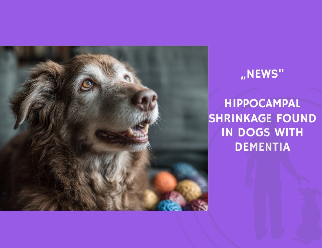
In a retrospective study led by **Curtis Wells Dewey** and colleagues, researchers analyzed MRI scans of **42 aging dogs (≥ 9 years old)**—including **16 dogs with diagnosed CCD** and **26 aging controls**—to determine whether CCD is associated with structural brain changes. The study was published on bioRxiv, a preprint server for biological research.
Using Mimics® software to calculate **total hippocampal and brain volumes**, the researchers found that dogs with CCD had a **significantly smaller hippocampal volume** relative to their total brain size (p = 0.04). The hippocampus is a brain region critical for **memory and spatial navigation**, and atrophy in this area is a hallmark of **Alzheimer’s disease in humans**.
This finding supports the idea that **canine cognitive dysfunction may serve as a natural model for human dementia**, enabling researchers to explore disease mechanisms and test potential therapies. In particular, **hippocampal-targeted interventions**, such as cognitive enrichment and pharmacological strategies, may benefit both dogs and humans.
Importantly, this study provides veterinarians and dog owners with **MRI-based evidence** that cognitive decline in senior dogs is not only behavioral but also **structurally visible in the brain**. Early recognition of CCD symptoms—such as disorientation, disrupted sleep, and changes in social interaction—may allow for timely intervention to slow neurodegeneration.
Source: Curtis Wells Dewey, M. Rishniw, S. Platt, K. R. Robinson, J. Sackman, M. O’Donnell. Published on bioRxiv, January 27, 2020. https://doi.org/10.1101/2020.01.27.921742