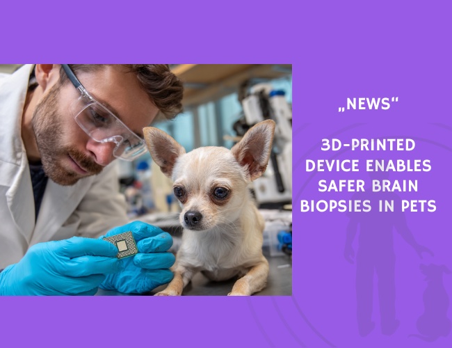
A team led by **Sabrina Fert and colleagues** has pioneered an innovative method to obtain brain biopsies in small animals using a **3D-printed stereotactic device** tailored to each patient’s skull and dental anatomy. The study, published in the American Journal of Veterinary Research, presents a breakthrough in neurosurgical precision and safety for companion animals.
The study involved **five clinical patients**—four dogs and one cat—with suspected brain lesions. MRI and CT imaging data were used to plan trajectories for the biopsy, and a custom 3D model was designed using **maxillary dental impressions** to stabilize the patient’s head. The printed device, mounted with biopsy guidance ports, allowed precise targeting of brain lesions.
As a proof of concept, the device was first tested on two cadaver heads before being applied in live animals. In **80% of the cases**, biopsy samples yielded a definitive histopathologic diagnosis, including **astrocytoma**, **meningioma**, **choroid plexus tumor**, and **chronic meningoencephalitis**. One case that initially yielded no diagnostic tissue was later confirmed as **low-grade oligodendroglioma** postmortem.
Importantly, all animals recovered well and were **discharged within 24 hours**, with no complications reported. The authors emphasize the system’s low cost, broad applicability to different skull shapes and lesion types, and potential to guide future treatment and prognosis with greater confidence.
This novel approach could revolutionize **neurologic diagnostics** in veterinary medicine, offering a minimally invasive and accessible option for accurately identifying brain pathologies that previously required more invasive methods or remained undiagnosed.
Source: S. Fert, H. Frankar, A. Dussaux, C. Berthomé, S. Chantrel, L. Cauzinille. Published in American Journal of Veterinary Research, July 2, 2024, Pages 1–10.