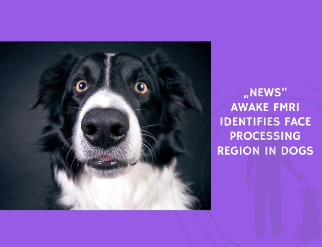
Dogs show remarkable sensitivity to human social cues, including eye contact, facial expressions, and gaze direction. While behavioral studies have demonstrated that dogs can visually discriminate and recognize faces, direct evidence for specialized cortical mechanisms supporting face perception had been lacking.
In this study, six dogs were trained to remain motionless in an MRI scanner while fully awake and unrestrained, an approach that avoids confounds associated with anesthesia. Using functional MRI, researchers measured neural responses while dogs viewed short movie clips of human faces and everyday objects.
The results revealed a region in the canine temporal cortex that responded significantly more to faces than to objects. This candidate dog face area was then tested using a new stimulus set. The region showed equally strong responses to human and dog faces, and significantly weaker responses to inanimate objects.
Importantly, this pattern of selectivity was not observed in primary visual cortex, indicating that the effect cannot be explained by low-level visual features such as contrast, shape, or motion. Instead, the findings support the presence of higher-order neural machinery dedicated to face processing.
These results provide the first neuroimaging evidence for a face-selective cortical region in dogs and demonstrate that specialized face-processing systems are not unique to primates. The findings suggest that convergent or conserved neural solutions for social perception may have evolved across distantly related mammalian lineages.
By identifying a neural substrate for face perception, this study offers a mechanistic explanation for dogs’ exceptional ability to attend to and interpret human facial cues, reinforcing the view of dogs as highly adapted social partners in the human environment.
Source: Dilks, D. D., Cook, P., Weiller, S. K., Berns, H. P., Spivak, M., & Berns, G. S. (2015). Awake fMRI reveals a specialized region in dog temporal cortex for face processing. :contentReference[oaicite:1]{index=1}.