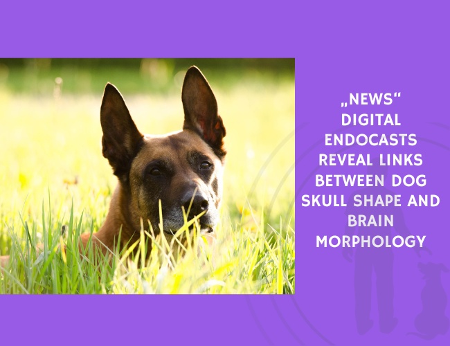
Study Chiang Mai, Thailand, December 24, 2025 – New digital canine endocasts visualize how differences in skull proportions reflect underlying brain morphology.
Digital endocasting, enabled through high-resolution computed tomography (CT), provides a unique window into canine brain morphology without requiring soft-tissue preservation. In this study, researchers generated detailed digital endocasts from skulls of 24 dog breeds alongside 4 wild canids to document a reproducible methodology for anatomical visualization and to explore how skull shape corresponds with the surface areas of major cerebral regions.
Endocasts were produced using CT scans at 0.323 mm × 0.322 mm × 0.6 mm resolution. Imaging data were segmented and refined using 3D Slicer and Autodesk Meshmixer, and interactive 3D PDFs and physical 3D prints were created to support teaching and comparative anatomical analysis. This approach allowed clear visualization of the prepiriform rhinencephalic region, prefrontal region, and non-prefrontal cerebral convexity.
Analyses showed that as the skull index increased—indicating broader, shorter skulls—the rhinencephalic area ratio decreased. This finding suggests that brachycephalic skull shapes may be associated with reduced surface proportion of olfactory-related structures, whereas longer-snouted breeds retain larger proportional areas in these regions.
The study demonstrates how digital endocasts can clarify the relationship between skull form and brain morphology, enabling future work linking neuroanatomical variation to behavioral traits and cognitive specialization in domestic dogs. These methods offer valuable tools for researchers, educators, and comparative anatomists.
Source: Czeibert, K., Sommese, A., Petneházy, Ö., Csörgő, T., & Kubinyi, E. (2020). Digital Endocasting in Comparative Canine Brain Morphology. Frontiers in Veterinary Science. https://doi.org/10.3389/fvets.2020.583945